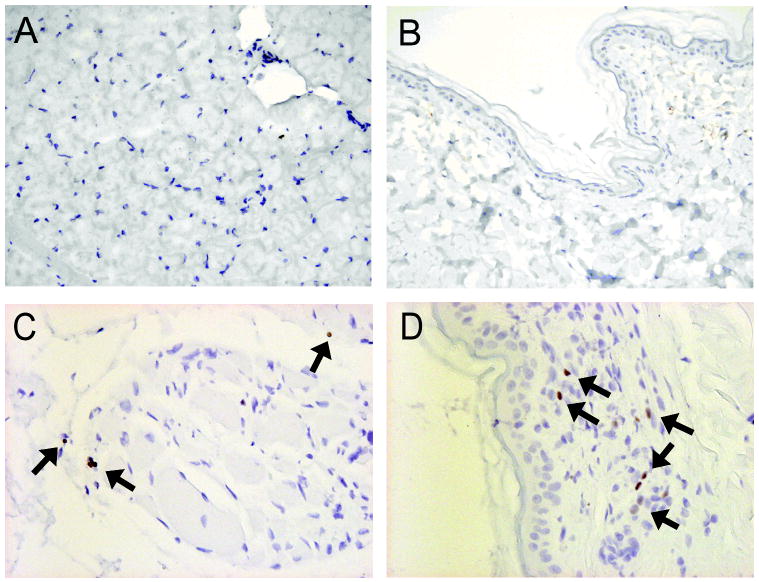
Figure 4. FoxP3+ staining of muscle and skin biopsies from untreated dogs, bone marrow donor dogs and vascularized composite allografts from mixed chimeric recipients.
Representative muscle (A) and skin (B) biopsies from an untreated dog showed no areas of FoxP3+ staining (200x magnification. In contrast, representative muscle (C) and skin (D) biopsies taken from the composite tissue allograft of mixed chimeric recipients (G767 and G969) reveal increased FoxP3+ staining with limited cellular infiltrate (Arrowheads; 400x magnification).

