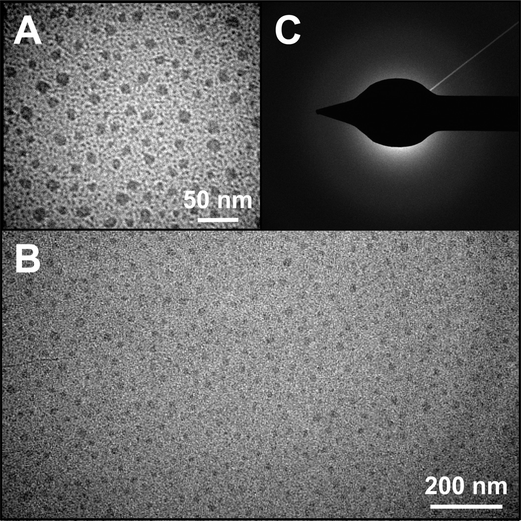
Figure 3.
TEM analysis of nanoparticles produced in the presence of TrxA::PA44. High (A) and low resolution (B) TEM images show that the characteristic size and polydispersity of CaP nanoparticles obtained under our experimental conditions. Selected area electron diffraction (SAED) patterns (C) were diffuse with no distinguishable spots or rings, suggesting that the mineral is amorphous calcium phosphate (ACP).

