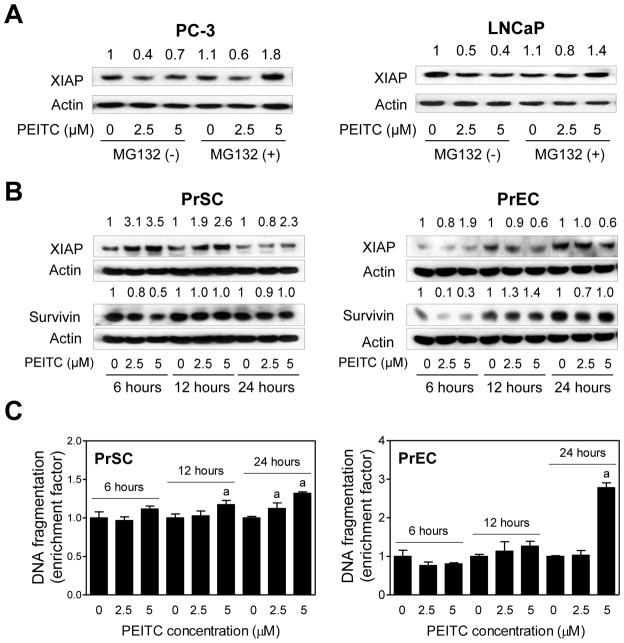
Fig. 2.
Normal prostate stromal and epithelial cells are relatively resistant to PEITC-induced apoptosis. A: Immunoblot for XIAP protein using lysates from PC-3 or LNCaP treated for 6 hours with DMSO (control) or the indicated concentrations of PEITC in the absence or presence of MG132 (1 μM for PC-3 and 5 μM for LNCaP). Cells were treated with MG132 for 2 hours and then exposed to DMSO or PEITC for an additional 6 hours with or without MG132. Blots were stripped and reprobed with anti-actin antibody to correct for differences in protein loading. Numbers on top of band represent change in XIAP protein level reletaive to DMSO-treated cells in the absence of MG132. B: Immunoblot for XIAP and Survivin proteins using lysates from PrSC and PrEC treated for specified time periods with DMSO (control) or the indicated concentrations of PEITC. Blots were stripped and reprobed with anti-actin antibody to correct for differences in protein loading. Numbers on top of bands are fold changes in expression level relative to the corresponding control. C: Quantitation of histone-associated DNA fragment release into the cytosol in PrSC and PrEC treated for specified time points with DMSO or the indicated concentrations of PEITC. Results shown are enrichment relative to corresponding DMSO-treated control. Results are mean ±SD (n=3). Significantly different (P< 0.05) compared with acorresponding DMSO-treated control by one-way ANOVA followed by Dunnett’s test. Similar results were observed in replicate experiments.

