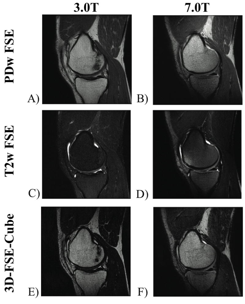
Figure 2.

Sagittal knee protocol images acquired at 3.0T and 7.0T. The 7.0T images have higher resolution, while maintaining similar fluid-to-cartilage contrast at each magnetic field strength with in-plane resolution of A) 0.29mm × 0.47mm, B) 0.15mm × 0.20 mm, (a 4.8x reduction in voxel volume at 7.0T). C) 0.39mm × 0.47mm, D) 0.20mm × 0.29mm, (a 3.2x reduction in voxel volume at 7.0T). E) 0.50mm × 0.59mm, F) 0.39mm × 0.59mm, (a 1.3x reduction in voxel volume at 7.0T).
