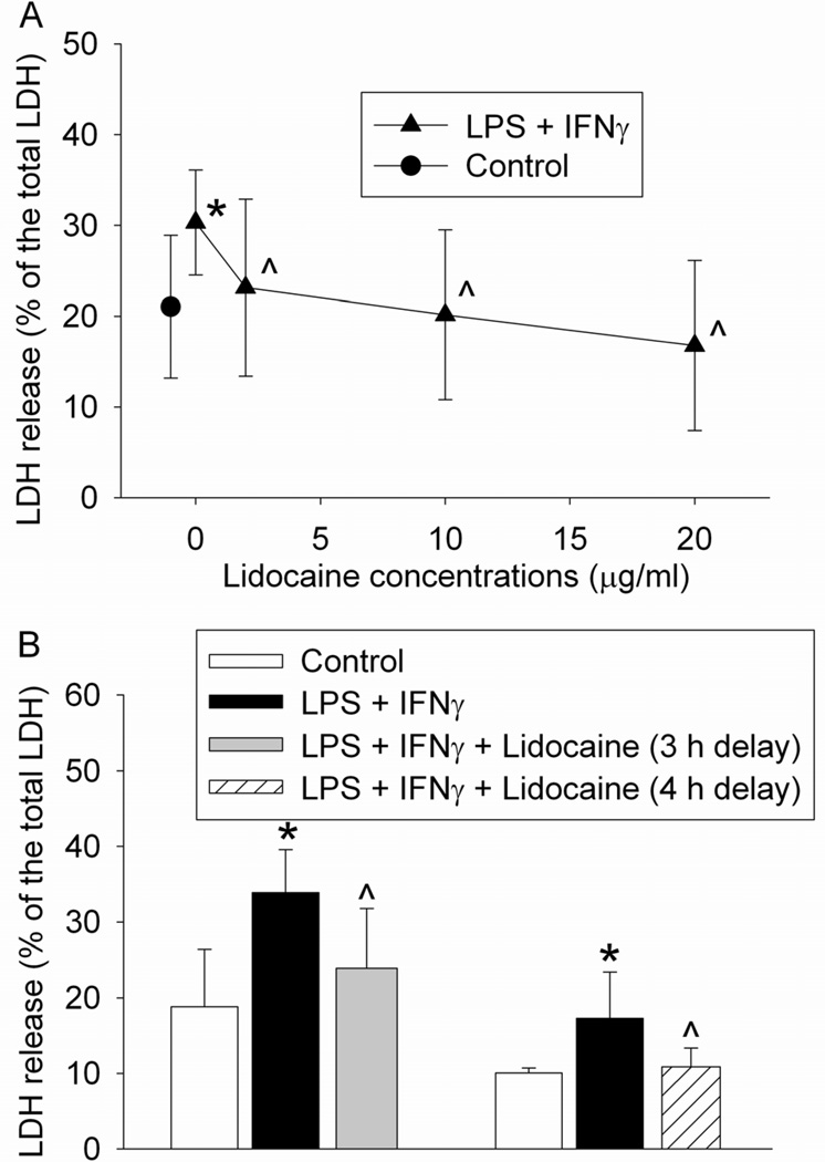
Fig. 2.
Protective effects of lidocaine on lipopolysaccharide (LPS) and interferon γ (IFNγ)-induced cell injury. (A) Dose-response of the lidocaine effects. The mouse C8-B4 microglial cells were incubated with 1 µg/ml LPS and 10 U/ml IFNγ for 24 h. Cells were exposed to 2, 10 or 20 µg/ml lidocaine for 1 h at 2 h after the initiation of the LPS and IFNγ stimulation. Cell injury was quantified by lactate dehydrogenase (LDH) release assay. Results are mean ± S.D. (n = 15 – 18). (B) Time-window of delayed isoflurane treatment. The mouse C8-B4 microglial cells were incubated with 1 µg/ml LPS and 10 U/ml IFNγ for 24 h. Cells were exposed to 20 µg/ml lidocaine for 1 h at 3 or 4 h after the initiation of the LPS and IFNγ stimulation. Results are mean ± S.D. (n = 6 – 17). * P < 0.05 compared to control. ^ P < 0.05 compared to LPS plus IFNγ only.

