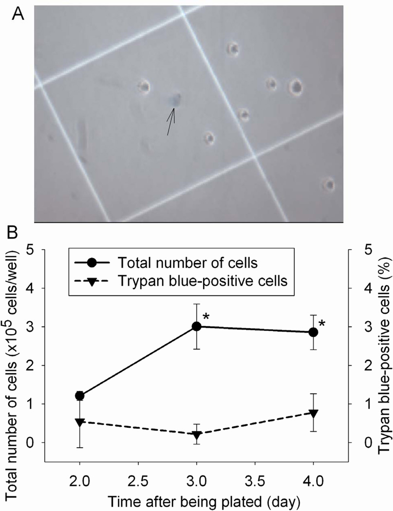
Fig. 5.
Cell viability under control condition. The viability of mouse C8-B4 microglial cells was evaluated by the trypan blue exclusion test at various times after they were plated. A representative microscopic view is presented in panel A and the pooled results are in panel B. The arrow in panel A indicates a trypan blue-positive cell. Results are means ± S.D. (n = 6). * P < 0.05 compared with the corresponding value at 1 day after the cells were plated.

