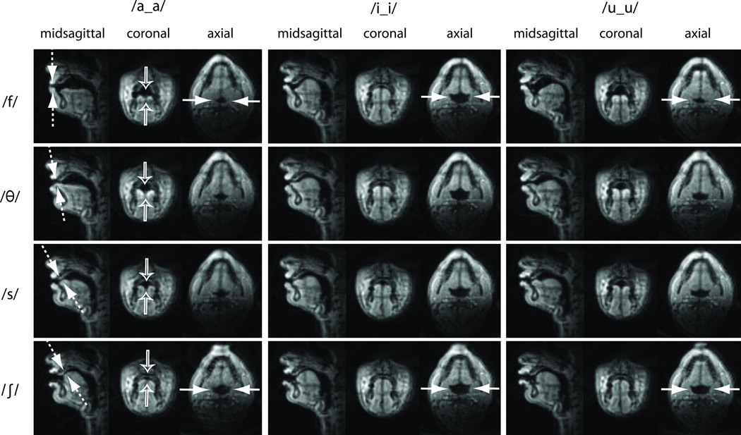
Figure 5.
Three-slice frames captured from Subject 2 using Protocol 2 during intervocalic voiceless fricative productions. Top row: labiodental fricative; 2nd row: dental fricative; 3rd row: alveolar fricative; Bottom row: post-alveolar fricative. Each scan plane provides unique vocal tract shaping information that is not available from the other scan planes. For example, the midsagittal slice shows the constriction locations (see the dashed arrows), the coronal slice shows the degree of tongue grooving/doming (see the hollow arrows), and the axial slice shows the degree of aperture and shaping of the pharyngeal airway (see the solid arrows). See also Movie 2 in the supplementary material.

