Figure 2.
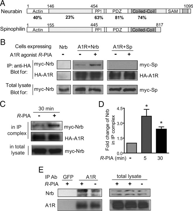
Agonist exposure specifically promotes interaction of the A1R with neurabin (Nrb) but not its homolog spinophilin in intact cells. A, Sequence homology between domains in neurabin and spinophilin. B, Interaction of the A1R with neurabin (left) or spinophilin (right) in cells treated with or without A1R agonist. Cells expressing neurabin alone or coexpressing HA-A1R with Myc-Nrb or Myc-Sp were stimulated with or without 1 μm R-PIA for 5 min, and cell lysates were subjected to immunoisolation assay using an antibody against HA. C, The A1R–neurabin interaction detected with prolonged R-PIA treatment for 30 min. Cells coexpressing HA-A1R and Myc-Nrb were stimulated with 1 μm R-PIA or vehicle for 30 min, and cell lysates were subjected to immunoisolation assay. D, Quantitation of A1R–neurabin interaction representing three to six independent coimmunoisolation experiments. Data are expressed as the fold change of neurabin in complex with the A1R over no stimulation control (defined as onefold). Values are given as the mean ± SEM; *p < 0.05, R-PIA stimulated versus control. E, Endogenous interaction between neurabin and A1R in mouse brain. Mice were given intraperitoneal injections of saline or R-PIA (1 mg/kg), with whole brains isolated and homogenized 30 min postinjection. Detergent-solubilized fractions were then prepared and subjected to coimmunoisolation using equal concentrations of an antibody against either GFP (negative control) or the A1R. Representative blots from three independent experiments are shown. A degradation product of the endogenous neurabin was also detected.

