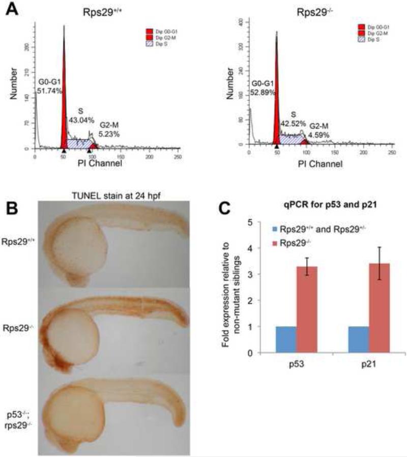
Figure 3. Apoptosis is induced in rps29-/- embryos.
(A) Cell cycle analysis. Propidium iodide was used to generate a cell cycle profile of rps29 homozygous mutants and their heterozygous and wildtype siblings at 24 hpf.
(B) TUNEL staining. TUNEL staining was performed on 24 hpf embryos from a rps29+/- incross, as well as embryos from a p53; rps29+/- incross.
(C) RT-PCR for p53 and p21. Data shown are fold changes of expression in rps29-/- mutant embryos compared to their heterozygous and wildtype siblings at 24 hpf.

