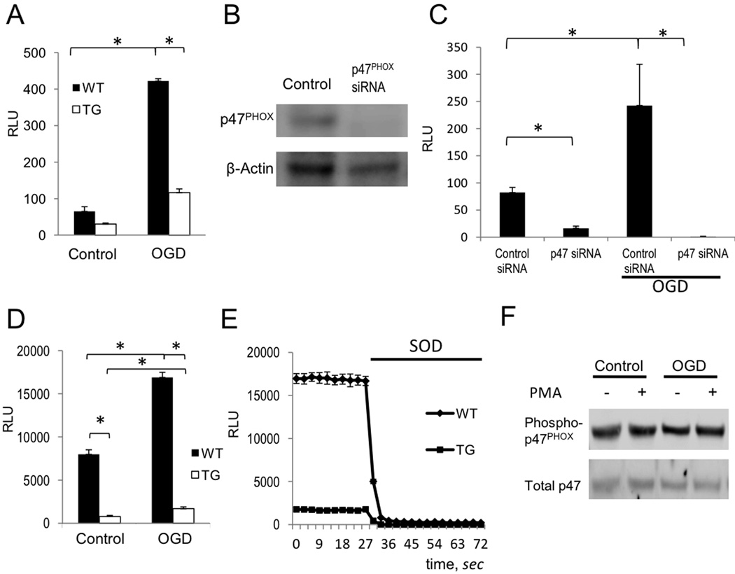
Figure 5. ROS production by PHOX in astrocytes is regulated by NF-κB.
(A) WT and TG astrocytes were assayed for superoxide production by Diogenes luminescence in control cultures and 24 hours after OGD challenge (OGD) (RLU = relative light units; n=5, p<0.05). (B) Transfection of p47PHOX siRNA reduces p47PHOX protein levels. Cell lysates were analyzed by western blot 24 hours post-transfection. (C) ROS production in live astrocytes was detected by Diogenes chemiluminescence 24 hours following OGD in non-targeting control siRNA (control) and p47PHOXsiRNA transfected cells. Astrocytes were transfected with siRNA 24 hours prior to OGD treatment. (n=5, *p<0.01). (D) Analysis of ROS production in WT and TG astrocytes 20 minutes after addition of PMA (1ng/ml) in control cultures and 24 hours after OGD (n=5, *p<0.01). (E) Representative recordings of ROS production from (D) in WT vs TG cells. Chemiluminesence was quenched by the addition of SOD (20U/ml). (F) Analysis of p47PHOX phosphorylation status by western blot in primary astrocyte extracts. OGD challenged and control astrocytes were treated with DMSO vehicle or PMA (1ng/ml) for 20 minutes prior to cell lysis.

