
Figure 1.
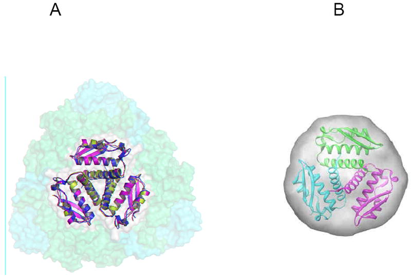
(A) Overlay of the γ subunits from Mtb (PDB code 2FVH - green), Klebsiella aerogenes (PDB code 2KAU – cyan) with 1.13Å RMSD, and Bacillus pasteurii (PDB code 1UBP- magenta) with 0.968Å RMSD superimposed onto the K. aerogenes urease trimer of trimers in semi-transparent orange (α) and yellow (β) surfaces highlighting the centralized location of the γ subunits in the urease complexes. (B) The envelope is calculated from the average of 16 GASBOR runs with P3 symmetry and 100 residues. The figures are reproduced from Reference 34.