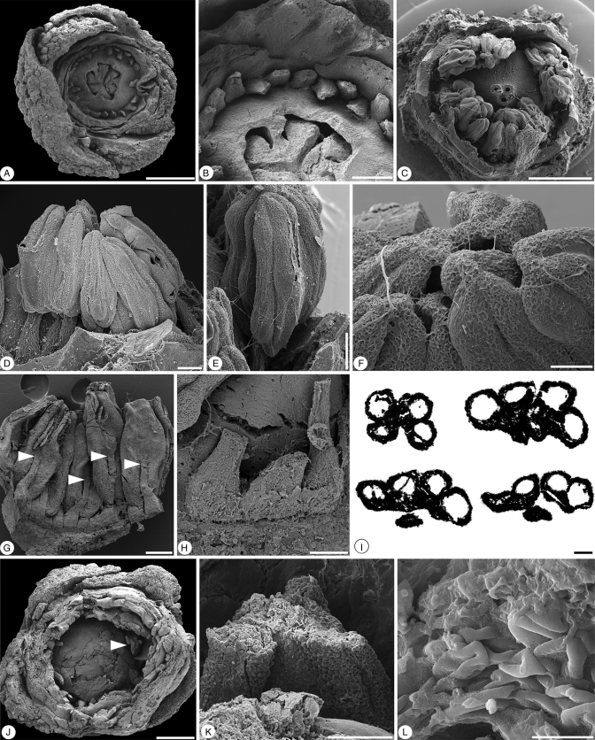
Fig. 3.
Glandulocalyx upatoiensis. Morphological details of stamens and pollen grains. (A) Apical view of damaged flower with part of perianth, anthers and top of ovary broken off, showing bases of stamen filaments arranged in a single series around the trilocular ovary with protruding-diffuse placentae; PP55156. (B) Detail of filament bases and protruding-diffuse placentae from the specimen in A; PP55156. (C) Apical view of the specimen in Fig. 1G–I dissected to reveal androecium and apex of gynoecium; PP55155. (D) Lateral view of the specimen in (C) showing stamens with filament and extrorse anther; PP55155. (E) Lateral view of stamen from specimen in (C); PP55155. (F) Apices of anthers from the specimen in (C) showing weakly developed connective tips; PP55155. (G) Adaxial view of group of stamens dissected from the specimen in Fig. 1F showing ventral (adaxial) anther attachment (arrows); PP55154. (H) Detail of broad filament bases; same specimen as in (G); PP55154. (I) Series of transverse sections through an anther of the specimen in Fig. 1D; ventral (adaxial) side facing downward; first section (upper left) showing the tetrasporangiate structure of the anther; second section (upper right) at the level of the joint between anther and filament; third section (bottom left) at the level below the joint of anther and filament with two theca united by a weakly developed connective; fourth section (bottom right) at the lower-most level where the theca are free from each other (sagittate anther); PP55160. (J) Apical view of broken, almost mature, floral bud; the arrow indicates partly preserved anther with in situ pollen shown in (K) and (L); PP55157. (K) Detail of broken anther with in situ pollen from specimen in J; PP55157. (L) In situ triaperturate pollen from the specimen in (J); PP55157. Scale bars (A, C, J) = 500 µm; (B, G) = 200 µm; (D, E, H, K) = 100 µm; (F, I, L) = 50 µm.

