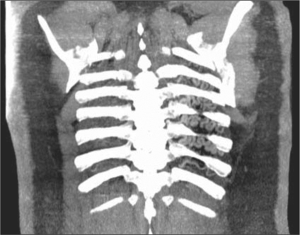
Figure 3.
Transaxial CT of the chest with intravenous contrast in a lung window shows distal branches of the left pulmonary artery that inappropriately communicate across the pleura with intercostal arteries. Once again, no proximal left pulmonary artery is noted. The left lung is hypoattenuating compared to a plethoric right lung.

