Figure 1.
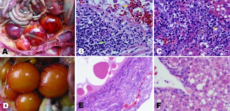
Pathologic changes in diseased Pekin ducks. A) Ovary with hyperemia, hemorrhage, and distortion. B) Ovary with hemorrhage (gold arrow), macrophage and lymphocyte infiltration and hyperplasia (green arrow). C) Liver with interstitial inflammation in the portal area (gold arrow). D and E) Ovaries from healthy ducks. F) Liver from healthy duck. A, C) Original magnification ×40; B, C, E, F) scale bars = 90 μm.

