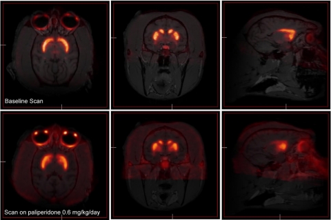
Fig. 5.
Sample [18F]fallypride PET scans coregistered with the animal's structural MRI scan are shown. An off-drug scan is shown in the top three images above, and the bottom three images depict a scan of an animal while the animal was receiving paliperidone. Three representative images are shown from each scan in the horizontal, coronal, and sagittal planes (from left to right). In both cases strong labeling in the caudate and putamen is easily identified.

