Figure 3.
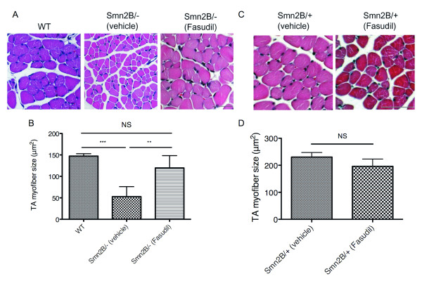
Fasudil increases tibialis anterior (TA) myofiber size. TA muscles were isolated from post-natal (P) day 21 untreated wild type (WT) (n = 3), vehicle-treated Smn2B/+ (n = 3), fasudil-treated Smn2B/+ (n = 3), vehicle-treated Smn2B/- (n = 6) and fasudil-treated Smn2B/- (n = 6) mice. (A) Representative images of cross-sections of WT, or vehicle- and fasudil-treated Smn2B/- TA muscles stained with hematoxylin and eosin. Scale bar = 50 μm. (B) Quantification shows that fasudil-treated Smn2B/- TA muscles display significantly larger myofibers than vehicle-treated Smn2B/- mice. (**P < 0.01; ***P < 0.001; NS = not significant; data are mean +/- s.d.). (C) Representative images of cross-sections of vehicle- and fasudil-treated Smn2B/+ TA muscles stained with hematoxylin and eosin. Scale bar = 50 μm. (D) Quantification shows that fasudil does not significantly increase the myofiber size of Smn2B/+ normal mice (NS = not significant; data are mean +/- s.d.).

