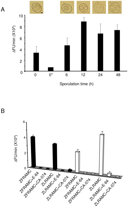
Figure 2. Biochemical activities of CPs throughout sporulation.
(A) Activities detected on Z-FR-AMC. Lysates of oocysts (1 mg/ml) taken at 0, 6, 12, 24 and 48 h after the beginning of sporulation were incubated with the substrate Z-FR-AMC (10 µM). The morphology of oocysts (under light microscopy) throughout the course of sporulation is shown. The scale bar represents 2 µm. (B) Activities detected on Z-FR-AMC and Z-LR-AMC in presence of the global cysteine protease inhibitor E-64 or the human cathepsin B specific inhibitor CA-074. Lysates (1 mg/ml) of oocysts taken at 0 h (black bars) and 48 h (white bars) after commencement of sporulation were pre-incubated with the inhibitors before adding the substrates. The data represents two independent experiments.

