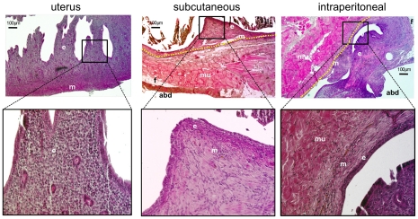
Figure 3. Endometriosis was confirmed with histology.
Haematoxylin and eosin staining was used to characterise histologically the lesions. ‘e’ indicates endometrial tissue; ‘m’ = myometrium. Muscle fibres and fat tissue of the abdominal wall are indicated by: ‘mu’ = muscle; ‘f’ = fat; ‘abd’ = abdominal cavity. The border between endometriosis and the abdominal wall is indicated by the yellow-dashed line.

