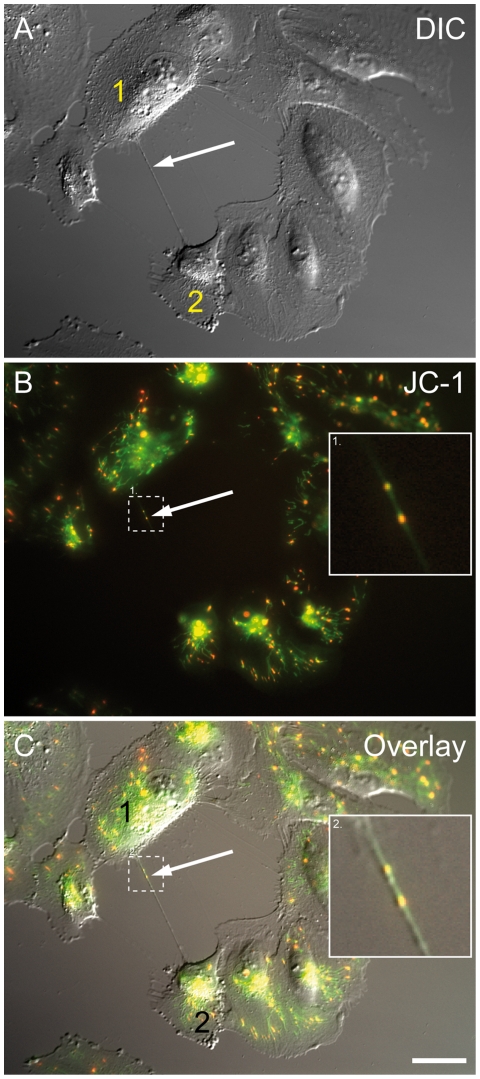
Figure 6. ARPE-19 cells connected by a nanotube containing mitochondria.
. (A) The bright field image shows two ARPE-19 cells connected by a membrane nanotube (arrow). (B) The corresponding fluorescence image of (A) shows JC-1 labelled mitochondria of cells (arrow). (C) The overlay of (A) and (B) shows the co-localization of nanotube with fluorescent labelled mitochondria (enlarged box). Scale bar, 20 µm.

