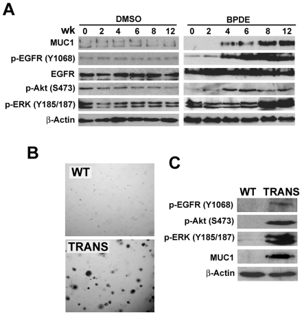
Figure 3. Chronic BPDE exposure activates Akt and ERK through EGFR in human bronchial epithelial cells.
A, Induction of MUC1 expression and EGFR-, Akt- and ERK-activation in HBEC-2 cells by BPDE. Specifically, HBEC-2 cells were treated with the vehicle DMSO or BPDE (0.1 µM) for the indicated weeks. Western blot was the same as in A. β-Actin was detected as a loading control. B, Increased MUC1 expression and EGFR-, Akt- and ERK-activation in transformed HBEC-2 cells by BPDE. HBEC-2 cells were treated with BPDE (0.1 µM) for 12 wk and then seeded in soft agar. Colonies were grown up for 3 wk in transformed cells (TRANS). Wild-type (WT, exposed to sham) HBEC-2 cells were as a negative control. Expression of MUC1 and activation of Akt and ERK were detected by Western blot in both WT and the transfected cells. β-Actin was detected as a loading control.

