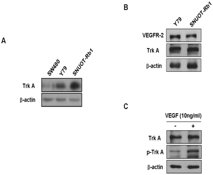
Figure 1. Activation of TrkA Induced by VEGF in Retinoblastoma Cells.
(A) Proteins of human retinoblastoma cell lines, Y79 and SNUOT-Rb1 [22] as well as a human colorectal cancer cell lines, SW480 were resolved on 12% SDS-PAGE and western blot analysis was performed using anti-TrkA antibody. β-actin was served as a loading control. Each figure is representative ones from three independent experiments. (B) Proteins of Y79 and SNUOT-Rb1 cells were resolved on 12% SDS-PAGE and western blot analysis was performed using anti-VEGFR-2 and anti-TrkA antibody. β-actin was served as a loading control. Each figure is representative ones from three independent experiments. (C) SNUOT-Rb1 cells were treated with 10 ng/ml VEGF. TrkA and phospho-TrkA were detected by Western blot analysis. β-actin was served as a loading control. Each figure is representative ones from three independent experiments.

