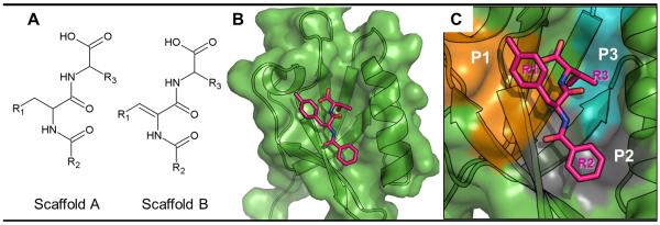
Figure 1.
Complex-structure-based pharmacophore model. (A) 2D structures of scaffolds A and B. (B) Complex structure of Dvl PDZ domain bound to scaffold-A compound 17. (C) Protein surface plotted in color to represent PDZ domain pockets P1 (orange), P2 (gray) and P3 (blue) that interact with R1, R2 and R3 fragments of 17 in the complex.

