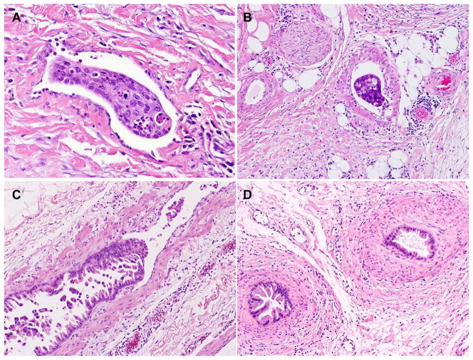
Figure 1.
Representative micrographs of tumor invasion into the lymphovascular space without muscle layer (non-muscular LVS, A) and tumor invasion into muscular vessels (B). Pancreatic ductal adenocarcinoma invades into and grows along the endothelial surface in a muscular vessel (C). D, Cross-sections of intravascular tumor growth shown in Figure C with smooth muscle layers wrapping around the tumor cells mimicking pancreatic intraepithelial neoplasia (PanIN). Hematoxylin & eosin stain, original magnifications: 200x

