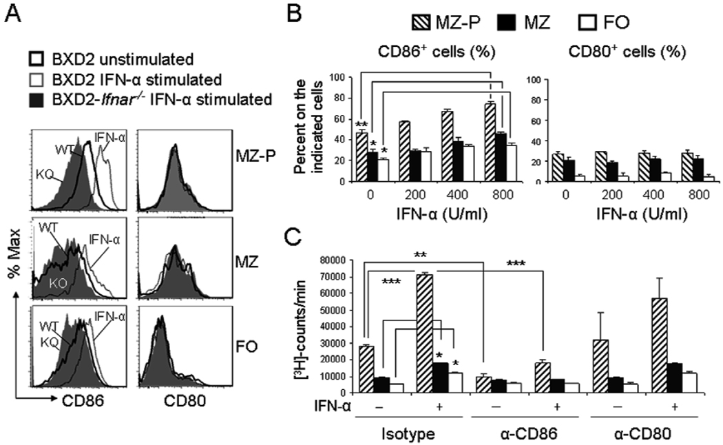
Figure 5.
IFN-α-induced costimulatory function of MZ-P B cells was suppressed by CD86 neutralization. A, FACS sorted MZ-P, MZ, and FO B cells from BXD2 and BXD2-Ifnar−/− mice were stimulated with medium or IFN-α (400 U/ml), as described in Materials and Methods. Results are shown as the representative FACS histograms showing the cell surface expression of CD86 and CD80 in the indicated population of B cells. B, Bar graphs showing the IFN-α-induced dose dependent induction of the expression of CD86 and CD80 on sorted MZ-P, MZ, and FO B cells. C, Tritiated thymidine proliferation assay of irradiated MZ-P, MZ, and FO B cells from BXD2 and BXD2-Ifnar−/− mice co-cultured with CD4 T cells and an agonistic anti-CD3 antibody for 48 hours. Sorted B cells were stimulated with or without IFN-α first as described in Materials and Methods. Cells were cultured in the presence of rat IgG2a κ isotype control (10 µg/ml), anti-CD86 (10 µg/ml), anti-CD80 (10 µg/ml). All results are shown as mean ± SEM (N=3 independent experiments from two mice per experiment, * p<0.05, **p<0.01, *** p<0.001 between the indicated comparison).

