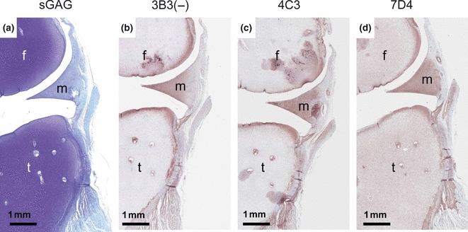
Figure 4.

Specific spatial expression of native CS/DS sulphation motif epitopes recognized by mAbs 3B3(−), 4C3 and 7D4 in a 12-week-old human foetal knee joint. Sections have been stained with Toluidine Blue [A] and immunostained with 3B3(−) [B], 4C3 [C] and 7D4 [D]. Note that Toluidine Blue staining identifies the presence of the major populations of CS GAG chains in the developing knee joint tissues. This Toluidine Blue staining is widespread and identifies the large number of GAGs present (mainly CS) throughout the early cartilage elements of the femur (f) and tibia (t) with weaker staining of the fibrocartilagenous meniscus (m) and other fibrous connective tissues of the joint. In contrast, immunostaining with the mAbs 3B3(−), 4C3 and 7D4 shows positive staining in very specific zones of the developing joint cartilage and meniscus that clearly delineate where the future articular cartilage and inner portions of the meniscus will eventually be. Close inspection of the three different mAb staining patterns shows that there are subtle differences in the mAb staining in the matrix or on the cells themselves indicating the specific spatial expression of these CS/DS sulphation epitopes.
