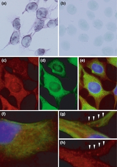
Figure 7.

In situ hybridization of fukutin and double-immunohistochemical staining of fukutin and vimentin in astrocytoma cells. In in situ hybridization, positive reaction is predominantly observed in the perikarya, but the cytoplasm including cytoplasmic processes is also stained (a). A negative control does not show positive reaction (b). In immunohistochemistry, fukutin (c) and vimentin (d) are widely distributed in the cytoplasm: (e) is a merged photograph of (c) and (d). Positive reaction of fukutin (red colour) is granular and that of vimentin (green colour) is filamentous (f–h). These two positive deposits appear to be partially co-localized or closely associated even in a peripheral part of cytoplasmic process (arrowheads).
