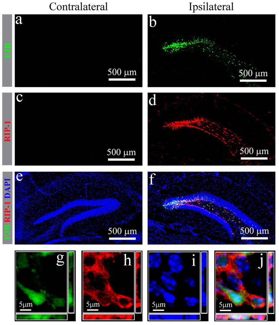
Figure 6.
Receptor interacting protein (RIP-1) expression in injured hippocampal granule neurons after moderate traumatic brain injury. (a–f) Distribution of injured neurons (a. b, green) and RIP-1 (c, d, red) in the contralateral (a, c, e) and ipsilateral (b, d, f) hippocampal dentate gyrus 24 hours post injury after combined Fluoro-Jade B (FJB) immunostaining. (g–j) A representative 3-dimensional image of the injured granule neurons (g) with expression of RIP-1 in the cytoplasm (h) and condensed nuclei (i) confirms colocalization (j) at the single-cell level.

