
Figure 2.
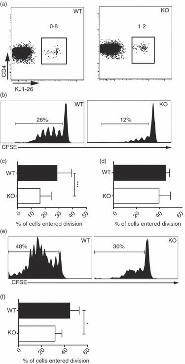
Reduced proliferation of CD4+ T cells in gut-associated lymphoid tissue (GALT) of CD47−/− mice after oral immunization. CFSE-labelled ovalbumin (OVA) -specific CD4+ T cells were transferred into CD47−/− or wild-type (WT) mice. Three days after oral immunization with (a) PBS, (b–c) OVA or (e, f) OVA + cholera toxin (CT) or (d) intravenous immunization with OVA, cells from (a–c) mesenteric lymph nodes (MLN), (d) spleen and (e, f) Peyer’s patches (PP) were analysed using flow cytometry. (b, e) Representative flow cytometric analysis of 7AAD− CD4+ KJ1-26+ cells. (c, d, f) Proliferation presented as frequency of T cells entering division from two to six pooled experiments. Each group consists of at least six individual mice. Error bars show SD.