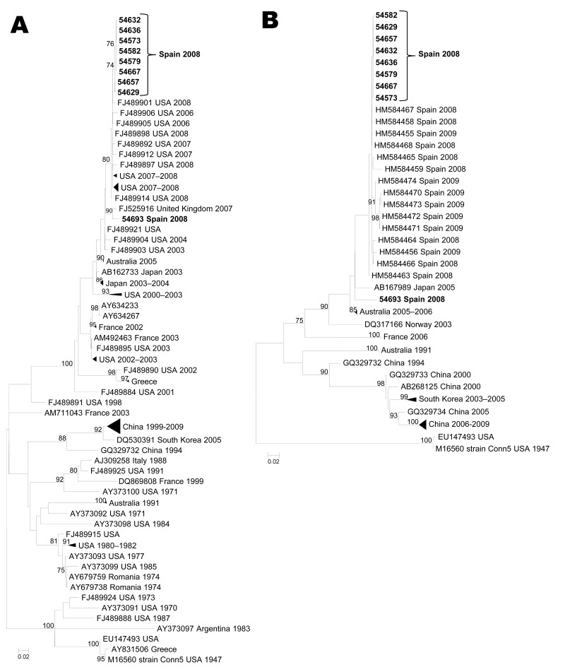
Figure 1.
Maximum-likelihood phylogenetic reconstructions for coxsackievirus B1 based on partial viral protein 1 sequences. A) 5′ partial coding region (93 sequences, 294 nt; B) 3′ partial coding region (49 sequences, 390 nt). Bootstrap values >75% are shown. Scale bars indicate number of substitutions per nucleotide position. Multiple strains from the same country sharing the same node were collapsed and shown as triangles with shape proportional to branch distances and number of sequences.

