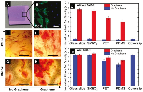
Figure 7.
Graphene substrate for osteogenic differentiation. A. Optical image of graphene-coated Si/SiO2 chip, showing the graphene boundary. B. Osteocalcin (OCN) marker showing bone cell formation on the same chip only on the graphene-coated area; C-D. Alizarin red quantification deriving from hMSCs grown for 15 days on substrates with/without graphene as well as with/ without BMP-2. E-H. PET substrate stained with alizarin red showing calcium deposits due to osteogenesis. 93 Reprinted with permission from ref.93. Copyright 2011 American Chemical Society.

