Fig. 3.
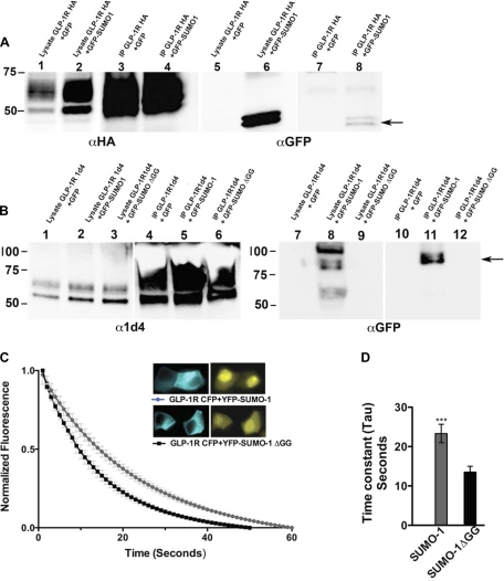
SUMO-1 binds both noncovalently and covalently to GLP-1R. A: MIN6 cells stably expressing GFP-SUMO-1 or GFP were transfected with hemagglutinin (HA)-tagged GLP-1 receptor (GLP-1R HA) and immunoprecipitated to detect protein interactions. To detect noncovalent interaction, cells were lysed without N-ethylmaleimide, immunoprecipitated with an anti-HA antibody, and detected with mouse GFP antibody. The arrow indicates an ∼40-kDa double band representing GFP-SUMO-1. Lane 1, cell lysate GLP 1RHA + GFP; lane 2, cell lysate GLP-1R HA + GFP-SUMO-1; lane 3, immunoprecipitation (IP) from lane 1; lane 4, IP from lane 2; lanes 1–4, blotted with rabbit anti-HA antibody; lanes 5–8 are similar to lanes 1–4, blotted with mouse anti-GFP antibody. B: to detect covalent interaction, MIN6 cells stably expressing GFP-SUMO-1 or GFP or conjugation-deficient GFP-SUMO-1ΔGG were transfected with GLP-1R 1d4, lysed, and immunopurified under denaturing conditions with anti-1d4 antibody, and the eluates were blotted with rabbit GFP antibody. Arrow indicates a ∼90-kDa immunoprecipitated band that represents GLP-1R covalently linked to GFP-SUMO-1 present only in the lane expressing GFP-SUMO-1 and GLP-1R 1d4. Lane 1, cell lysate GLP-1R 1d4 + GFP; lane 2, cell lysate GLP-1R 1d4 + GFP-SUMO-1; lane 3, GLP-1R 1d4 + GFP-SUMO-1ΔGG; lane 4, IP from lane 1; lane 5, IP from lane 2; lane 6, IP from lane 3; lanes 1–6 are blotted with mouse anti-1d4; lanes 7–12 are the same as lanes 1–6 and blotted with rabbit anti-GFP antibody. C: FRET analysis by donor bleaching between GLP-1R-cyan fluorescent protein (CFP) and yellow fluorescent protein (YFP)-SUMO-1 shows interaction between GLP-1R and SUMO. Graph shows normalized fluorescent decay curves in cells expressing GLP-1R-CFP and YFP-SUMO-1 (gray) or GLP-1R-CFP and YFP-SUMO-1ΔGG (black). Inset: image of representative cells expressing GLP-1R-CFP and YFP-SUMO-1 or YFP-SUMO-1ΔGG; n = 14–17 cells from multiple experiments. D: graph showing time constants (tau) of the decay curves. Cells expressing GLP-1R-CFP and YFP-SUMO-1 showed 1.7-fold increase in time constant compared with cells expressing GLP-1R-CFP and YFP-SUMO-1ΔGG, indicating protein-protein interaction between GLP-1R and SUMO-1 but not with conjugation-deficient SUMO-1. Error bars indicate means ± SD; n = 6. Student's t-test: ***P < 0.0007.

