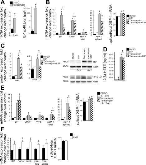
Fig. 3.
ER stress signals through IL-12 and is attenuated by the anti-inflammatory agent lysofylline (LSF). A–E: 3T3-L1 adipocytes were treated with 5 μM tunicamycin or DMSO solvent control for 24 h, pretreated for 2 h, and coincubated with the anti-inflammatory compound LSF (10 μM) or with 2 μg/ml IL-12p40 antibody (Ab). F: 3T3-L1 adipocytes were treated with 2.5 ng/ml mouse IL-12 cytokine for 24 h. mRNA (A, B, E, and F) and protein measurements (C) of IL-12p40, ER stress markers, and 12/15-LO were examined. IL-12p40 (A) and 12(S)-HETE (D) were measured in media cultured with treated 3T3-L1 adipocytes by ELISA. The mRNA measurements were done by RT-PCR and protein measurements done by Western blot analysis; the data were normalized to total actin, and the fold changes in expression were calculated relative to DMSO control. Representative Western blots are shown, and separate panels for each antibody represent the same exposure; however, samples for BiP and corresponding actin were run on different gels and clearly demarcated (C) (see materials and methods for quantitation). All data represent means ± SE; n = 3. Statistics were performed, comparing each treatment with control as well as tunicamycin with tunicamycin + LSF/Ab; only statistically significant results are shown. *P < 0.05, †P < 0.02, and #P < 0.001 vs. control unless otherwise indicated.

