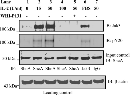
Fig. 2.
IL-2 promotes Jak3 interactions with ShcA at lower concentrations: HT-29 CL-19A cells were treated with 15-, 50-, and 100-U/ ml of IL-2 in the presence or absence of Jak3 inhibitor WHI-P131 for 15 min. Cells were lysed using lysis buffer and proteins in the lysates were estimated using BCA protein assay reagents. Equal amounts of ShcA proteins (Input control) were subjected to immunoprecipitation (IP) using Shc-A (test samples; lanes 1–5), Jak3 (positive control; lane 6), and IgG (negative control; lane 7) antibodies. IB, immunoblot. Western analysis of the SDS-PAGE separated protein complex was performed using a Jak3 and phospho-tyrosine (pY20) antibodies. Blots shown represent n = 3 experiments.

