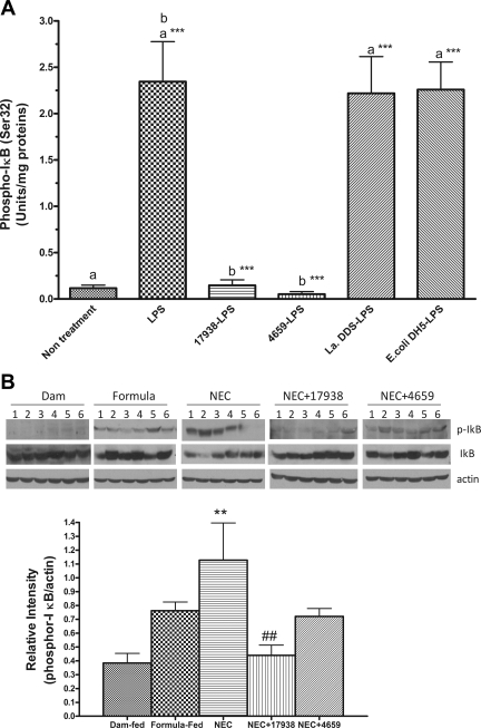
Fig. 5.
Phospho-IκB activation in the intestines of newborn rats in an ex vivo experiment (A) and in the NEC model (B). A: ileal tissues from each newborn rat (day of life 0) were collected in an antibiotic-free DMEM complete medium and immediately treated with probiotics (strain17938 or strain 4659) or control bacteria [Lactobacillus acidophilus DDS (La DDS) or Escherichia coli DH5α; 3–4 × 10⁷ CFU] for 4 h, washed, and subsequently treated with LPS (E. coli subtype 0111:B4), 100 μg/ml for 30 min. Tissues were washed with PBS and homogenized in a lysis buffer containing protease inhibitors. Tissue lysates were used to determine phospho-IκB activity by ELISA. Activity of phospho-IκB (Ser32) was expressed as units/ mg total protein in tissue lysates; n = 6 rats for each group. aGroups compared with no treatment; b17938-LPS or 4659-LPS compared with LPS. Strains 17938 and 4659 both inhibited IκB phosphorylation by > 90%: ***P < 0.001. B: phospho-IκB analysis in the ileum of rat pup with NEC as examined by Western blot analysis. Fifty micrograms of total protein were loaded and immunoblotted with anti-phospho-IκB and IκB antibodies, with β-actin as a loading control. Top: a typical blot; numbers represent number of animals examined in each group. Bottom: relative densitometric values are means ± SE; n = 6 for each groups. **P < 0.01, NEC vs. dam fed; ##P < 0.01, NEC + 17938 vs. NEC.

