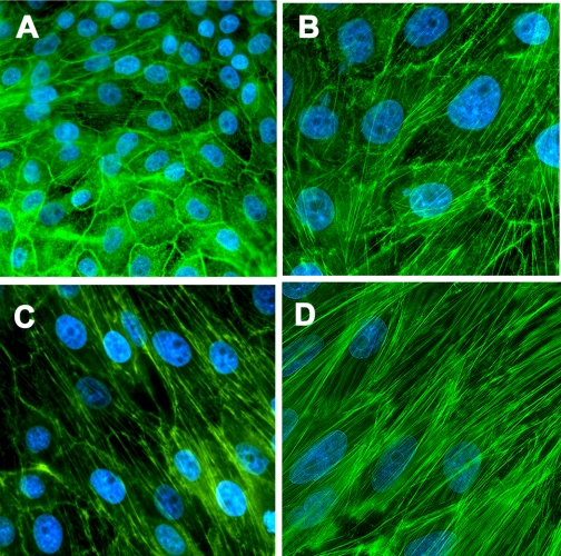
Fig. 3.
Chronic NIC exposure aggravates TGF-β1-induced stress fiber formation in a U-STAT3-dependent fashion in cultured renal proximal tubule cells. LLC-PK1 cells were treated with 10 ng/ml TGF-β1 for 3 days (B–D). In a set of experiments, cells were pretreated with 200 μM NIC (C and D) for 24 h before treatment with TGF-β1. Cells in D were infected with a Y705F-STAT3 adenovirus for 24 h before treatment. Cells were fixed and stained with Alexa Fluor 488-labeled phalloidin plus 4′,6-diamidino-2-phenylindole (DAPI; magnification ×400). Results are representative of 3 separate experiments.

