Fig. 4.
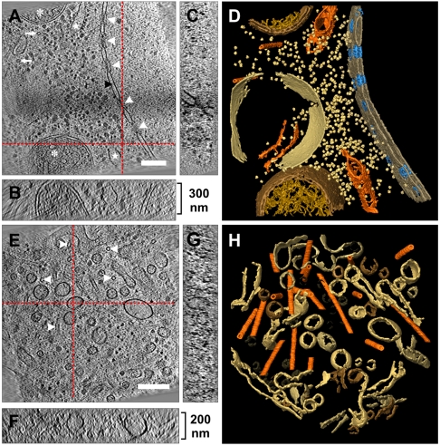
Cryoelectron tomograms of D. discoideum cells. (A) Slice through the x,y-plane of a tomographic reconstruction (area corresponds to the exposure region described in Fig. 3) showing the nuclear envelope (black arrowhead) with nuclear pore complexes (white arrowheads) separating cytoplasm from nucleoplasm. In the cytoplasm, parts of the rough endoplasmic reticulum (white stars), tubular mitochondria (asterisks) and microtubules (white arrows) can be recognized. (B and C) Displays the corresponding x,z and y,z-planes (sectional planes are indicated by the red dashed lines). The thickness of the lamella is approximately 300 nm. (D) Surface rendered visualization of the tomographic volume from (A), displaying nuclear envelope, endoplasmic reticulum, mitochondria, microtubules, vacuolar compartment, and putative ribosomes. (E) Tomographic slice along the x,y-plane showing a cytoplasmic volume traversed by several microtubules (white arrowheads). (F and G) Corresponding x,z and y,z-planes. The overall volume has a thickness of approximately 200 nm. (H) Surface rendered visualization of the tomographic volume from (E), showing microtubules (orange) traversing the volume in various directions. A dense network of interconnecting tubular structures and vesicles can be seen. [Scale bars, (A and E) 200 nm.]

