Fig. 5.
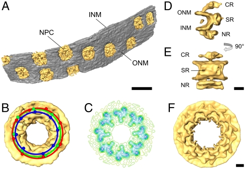
Structure of the nuclear pore complex from D. discoideum obtained by subtomogram averaging of the NPCs from the tomogram depicted in Fig. 4. (A) Surface representation of the segmented nuclear envelope (NPCs: yellow; nuclear envelope: gray). (B) Structure of the NPC obtained by averaging the three full pores in the tomogram, without imposing rotational symmetry. The red, green, and blue dots represent the positions of the protomers for each of the pores. Note that while all pores are round, there is a variation in their diameter (10%) and from the ideal 45 degrees dictated by eightfold symmetry. (C) Contour-line representation of a slice of the cytoplasmic ring of the NPC. (D and E) Views of a protomer of the NPC obtained from averaging all protomers in the pores depicted in (A) (D: side view; E: view from the central channel). (F) Structure of the NPC reconstructed by applying eightfold symmetry to the protomer depicted in (D and E). For clarity, the material in the central channel (approximately 50 nm) is omitted in (B to F). (B, C, F) view from the cytoplasm. NPC: nuclear pore complex; ONM: outer nuclear membrane; INM: inner nuclear membrane; CR: cytoplasmic ring; SR: spoke ring; NR: nuclear ring. [Scale bars: (A) 200 nm, (B–F) 25 nm.]

