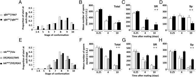
Fig. 1.
OA and TA do not contribute to early sperm storage events but are required for sperm depletion from storage. Sperm storage events are shown in OA-less females (A–D) and in OA/TA-less females (E–H). Distribution of uterine conformational stages, shown as proportions, at 35 min after the start of mating in tβhM18 (A) and tdc2RO54/Df(2R)42 mutant females (E) compared with their sibling controls (A: NM18 = 40, NFM7 = 50; E: NRO54/Df(2R)42 = 30, NGla = 30, NSM5 = 42). Total sperm stored in the SSOs (B and F), only in the seminal receptacle (C and G), and only in the spermathecae (D and H) in tβhM18 and tdc2RO54/Df(2R)42 mutant females and their sibling controls. Differences between/among female genotypes in the depletion of stored sperm over time were analyzed using two-factor ANOVA. The significance of the genotype factor, indicating differences in sperm depletion between/among mutant and control females, is reported in the figure as follows: ++ = P < 0.005 (an additional explanation of the statistical analysis is provided in Materials and Methods). Sp, spermathecae; SR, seminal receptacle. Sample sizes for sperm counts range from n = 7–20 (Table S1).

