Fig. 4.
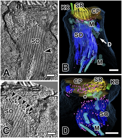
The striated organelle in cell 4, an extrastriolar type II hair cell, modeled by joining nine serial tomograms (total volume thickness = 4.5 μm). (A) A large portion of the SO is visualized in a single plane in SLICER mode, illustrating the planar nature of the SO in type II cells. Morphing of a thick bundle into a thin one can be observed (arrowhead). (B) A partial 3D reconstruction of the same hair cell. “D” in B refers to the view in D (i.e., from under the cuticular plate). (C) In another section from the same hair cell, note the dense spherical objects (arrowheads, depicted as pink spheres in B and D) aligned with many of the thin filaments along the margins of the SO. (D) View from below the cuticular plate: SRs are not connected to the SO, but may be associated with the dense spherical objects (pink spheres). Most mitochondria, except for those in close association with the SO, have been removed for clarity. (Scale bars, 0.5 μm.) CP, cuticular plate; KC, kinocilium; M, mitochondria; *, centriole.

