Fig. 7.
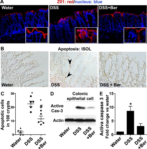
Berberine preserves intestinal barrier function in DSS-treated mice. Mice were treated as shown in Fig. 2. Paraffin-embedded colon tissues were used to determine zonula occludens-1 (ZO-1) distribution by immunohistochemistry using an anti-ZO-1 antibody and FITC-labeled secondary antibody and visualized by fluorescence microcopy (red staining). Nuclei were stained with 4,6-diamidino-2-phenylindole (DAPI; blue staining) (A). Apoptosis was detected using ApopTag In Situ Oligo Ligation (ISOL) staining. Apoptotic nuclei labeled with peroxidase were visualized using DIC microscopy (B). Arrows indicate ISOL-labeled apoptotic nuclei (brown in B). The number of apoptotic nuclei per 100 crypts is shown (C). Colonic epithelial cells were isolated from mice. Total cellular lysates were prepared for detecting caspase-3 (casp-3) activation by Western blot analysis (D). Blotting for actin was used as a protein loading control. Each lane represents epithelial cells from 1 mouse. The relative density of the activated caspase-3 band on Western blot was compared with the actin band in each group. The density ratio in the water group was set as 100%, and ratios of DSS and DSS+Ber were compared with that of the water group. Fold change of ratios is shown in E; n = at least 5 mice in each group. In C and E, *P < 0.05 compared with water groups.

