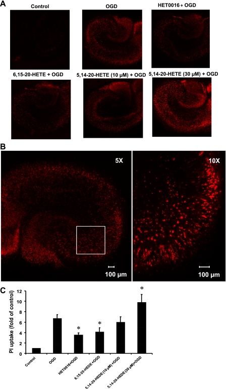
Fig. 2.
Effects of HET0016, 6,15-20-HEDE, and 5,14-20-HEDE on OGD- and reoxygenation-induced increases in propidium iodide (PI) uptake. Hippocampal slices were pretreated with HET0016 (10 μM), 6,15-20-HEDE (10 μM), 5,14-20-HEDE (10 and 30 μM), or vehicle for 30 min before OGD and throughout the experiment. Slices were exposed to 90 min of OGD followed by 2 h of reoxygenation. PI uptake was analyzed at the end of the reoxygenation period by confocal microscopy using a ×5 objective (flattened images of 200- to 300-μm z-stacks). A: representative PI fluorescence images indicating neuronal cell damage in the hippocampal slices. B: ×10 magnified image of a region (box) of the same hippocampal slice showing a close-up view of the cellular damage. C: quantification of cell death using total PI fluorescence. Results are expressed as fold increases over control. Values in C are means ± SE of 4 experiments/group. *P < 0.05 vs. the OGD group.

