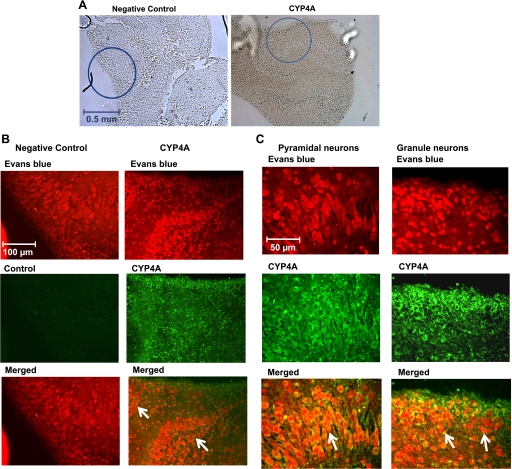
Fig. 4.
Immunohistochemistry for cytochrome P-450 (CYP)4A protein in organotypic hippocampal slices. Hippocampal sections were stained with CYP4A antibody followed by an Alexa fluor 488 secondary antibody (green). Sections were counterstained with 0.001% Evans blue to suppress autofluorescence and to allow cellular structures to be viewed using a rhodamine filter. Hippocampal sections incubated without primary antibody served as negative controls. A: low-magnification images (×5 objective: approximately ×50) of the hippocampal tissue showing the regions (circles) from where the images of immunohistochemical staining for CYP4A and negative control were taken. B: intermediate-magnification images (approximately ×250) of the hippocampal region showing that the CYP4A enzyme is widely expressed in the cell bodies of many neurons (merged images, indicated by arrows). C: high-magnification images (approximately ×500) showing CYP4A-positive neuronal cells in the hippocampal region showing that CYP4A protein is expressed in the cell bodies of both pyramidal and granule neurons (merged images, indicated by arrows).

