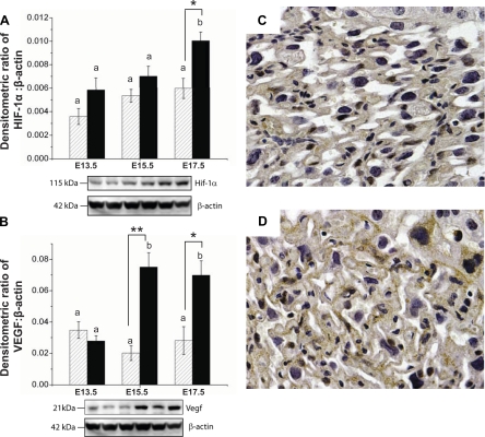
Fig. 3.
Potential factors driving vascular development in CD1 (hatched bars) and B6 (solid bars) mice. Western blots showed increased expression of hypoxia-inducible factor (HIF)-1α protein (A) in B6 placentas at E17.5 and VEGF protein (B) in B6 placentas at E15.5 and E17.5. The order of the lanes corresponds to the order of the bars on the graph. C and D: representative images of the CD1 (C) and B6 (D) placental labyrinth at E17.5 immunostained for HIF-1α. Sections from one placenta per litter from three CD1 dams and three B6 dams were examined. Staining in B6 mice was more pronounced. Western blot data are presented as means ± SE, where n = 4 dams per group per gestational age (tissue pooled from 3 placentas/litter). Two-way ANOVA was used to assess the effects of gestational age and strain. a,bDifferences with gestational age; *P < 0.05 and **P < 0.001, strain differences as determined by Tukey post hoc tests.

