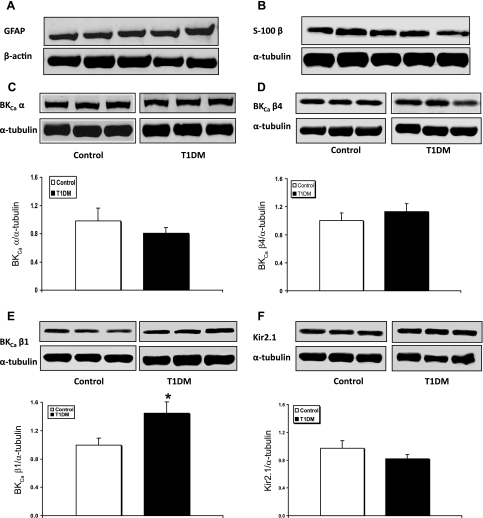
Fig. 3.
Western immunoblot protein analyses of GFAP, S-100 β, BKCa channel subunits, and Kir2.1 channels in samples obtained by stripping off pial tissue from the cerebral cortex surface in control and 4 mo diabetic (T1DM) rats. Those samples not only included pial vessels but also adherent elements of the underlying glia limitans, as revealed by the expression of astrocytic markers GFAP (A) and S-100 β (B). Also shown are representative blots and quantitative protein expression data for BKCa channel α-subunit (BKCaα) (C); BKCa channel β4-subunit (BKCaβ4) (D); BKCa channel β1-subunit (BKCaβ1) (E); and Kir2.1 channels (F) in control and T1DM rats. *P < 0.05; n = 6–7 in each group. Each graph shows the average of 3 independent blots. α-Tubulin or β-actin was used as a loading control. Ratios were normalized to the control's group average. The bands within a frame are contiguous, and splicing is shown by a white space between frames.

