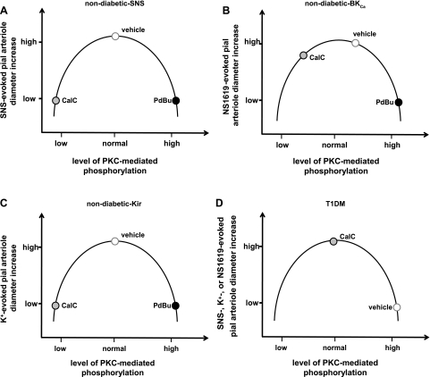
Fig. 6.
A–D: illustrations representing the postulated responses of pial arterioles to SNS, NS-1619, and K+ in control animals in the presence of PdBu-associated increases or CalC-induced decreases in the levels of PKC-mediated phosphorylation. For all 3 vasodilators, with the starting level of phosphorylation (vehicle) lying near the peak of the inverted U curve, PdBu-associated enhancement of phosphorylation will be accompanied by a repressed vasodilation (as the data in Fig. 5 indicate). Going in the other direction (CalC-associated diminished phosphorylation), vascular reactivity may also be reduced. That could apply to SNS- and K+-evoked responses (see Fig. 4, C and F). If one modestly shifts the level of baseline PKC-mediated phosphorylation to the right of the peak, then, during a CalC-associated leftward shift, one might observe no change in response, similar to what we observed for the BKCa opener NS-1619 (Fig. 4D). In diabetic (T1DM) rats (D), note that the baseline (vehicle) PKC-linked phosphorylation levels are depicted as being elevated, based upon the increased PKC activity measured in the glio-pial tissue (shown in Fig. 4B). In that case, a decrease in PKC-mediated phosphorylation levels (leftward shift along the curve) improved responses to all dilating stimuli.

