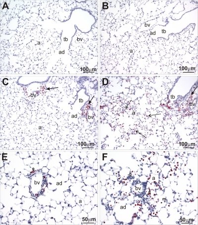
Fig. 3.
Major basic protein (MBP) immunohistochemistry. Lung sections from HIF-1α-sufficient (A, C, and E) and PN4 HIF-1α-deficient (B, D, and F) mice following sensitization and challenge with saline (A and B) or OVA (C, D, E, and F) were stained for MBP, an eosinophil-specific marker, via immunohistochemistry. Solid arrows indicate eosinophils located around terminal bronchioles and blood vessels, whereas dashed arrows indicate eosinophils located out in the parenchyma. a, Alveolus; bv, blood vessel; tb, terminal bronchiole; ad, alveolar duct.

