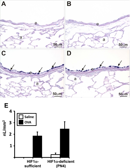
Fig. 4.
Alcian Blue-Periodic Acid Schiff (AB-PAS) staining and mucous quantification. Lung sections from saline (A and B) and ovalbumin (C and D) sensitized and challenged HIF-1α-sufficient (A and C) and PN4 HIF-1α-deficient (B and D) mice were stained with AB-PAS. Solid arrows indicate areas of positive AB-PAS staining. a, Alveolus; e, epithelium. Quantification of volume density of mucous (in nl/mm2) from AB-PAS immunohistochemistry from saline (white bars) and OVA (black bars) control and PN4 HIF-1α-deficient mice was performed as described in materials and methods (E).

