Fig. 6.
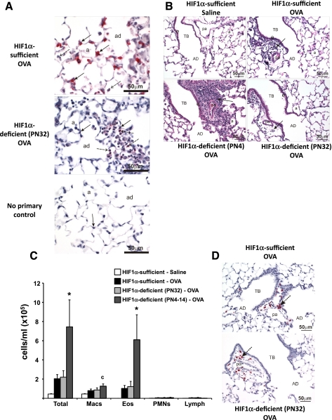
HIF-1α immunohistochemistry, hematoxylin and eosin (H&E), and BALF cellularity for PN32 and PN4–14 mice. HIF-1α immunohistochemistry of HIF-1α-sufficient (top) and PN32 HIF-1α-deficient (middle) mice after OVA sensitization and challenge is shown. No primary antibody control for HIF-1α from a HIF-1α-sufficient, saline-treated mouse is also pictured (bottom). Solid arrows indicate alveolar type II cells and dashed arrows indicate macrophages (A). Light photomicrographs of a centriacinar region from the lungs of HIF-1α-sufficient (top left and right) and HIF-1α-deficient PN4 (bottom left) and P32 (bottom right) mice treated with saline (top left) or OVA (top right and bottom left and right). Tissues were stained with H&E. Arrows indicate areas of cellular infiltrate (B). HIF-1α-sufficient (white and black bars), PN32 HIF-1α-deficient (light gray bars), and PN4–14 HIF-1α-deficient (dark gray bars) mice were sensitized/challenged with saline (white bars) or OVA (black, light gray, and dark gray bars). Total cells, as well as the numbers of macrophages (Macs), eosinophils (Eos), neutrophils (PMNs), and lymphocytes (lymph) were determined from BALF (C). Values are the mean ± SE (n ≥ 7). *Significantly different from all other groups P < 0.05; c, significantly different from saline treated. MBP immunohistochemistry performed on PN32 HIF-1α-deficient (bottom) verifying the lack of a significant increase in eosinophils in these OVA-treated lungs as compared with HIF-1α-sufficient (top) mice is shown. Arrows indicate similar areas of eosinophilic infiltration surrounding the pulmonary artery (D). AD, alveolar duct; a, alveolus; TB, terminal bronchiole; pa, associated pulmonary artery.

