
Figure 3. Selective reduction of mesoridazine affects cell viability.
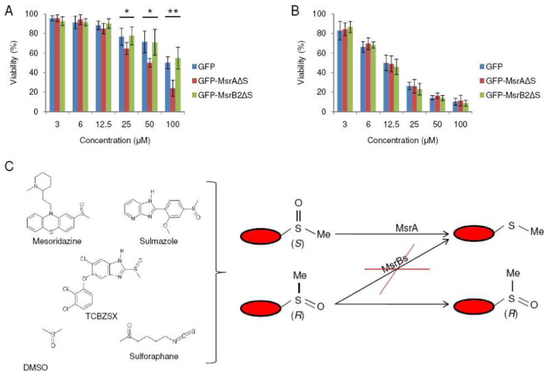
(A) Cell viability analysis of HEK 293 cells expressing GFP (control), GFP-fused mMsrAΔS, or GFP-fused mMsrB2ΔS following treatment of cells with mesoridazine (3, 6, 12.5, 25, 50, 100 μM) for 24 h. Constructs were designed to express proteins in the cytosol (where indicated by removing the signal peptide marked as ΔS). (B) Cell viability analysis of HEK 293 cells transfected with GFP (control), GFP-fused mMsrAΔS, or GFP-fused mMsrB2ΔS following treatment with thioridazine (3, 6, 12.5, 25, 50, 100 μM) for 24 h. Constructs were designed to express proteins in the cytosol (ΔS indicates that the signal peptide was removed). All measurements were repeated 6 times independently, and the data analyzed with a Student's t-test (**: p<0.01 and *: p<0.05). (C) An overall scheme of stereospecific reduction of methylsulfinyl-containing drugs.