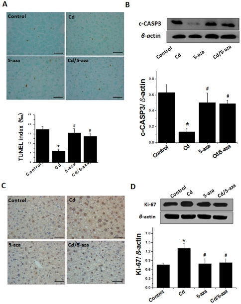
Figure 5. Apoptotic and cell proliferation effects of chronic exposure to Cd on mouse livers.
Liver samples were collected as described in Figure 3 for TUNEL staining with semi-quantitative evaluation (A). Apoptotic effect was further confirmed with cleaved-CASP3 (c-CASP3), detected by Western blot (B). Ki-67 measurement with immunohistochemical staining (C) and Western blot (D). The images for each group are the representative ones. Bar = 50 µm. Data was presented as mean ± SD (n = 10). * p<0.05 vs control; # p<0.05 vs Cd group.

